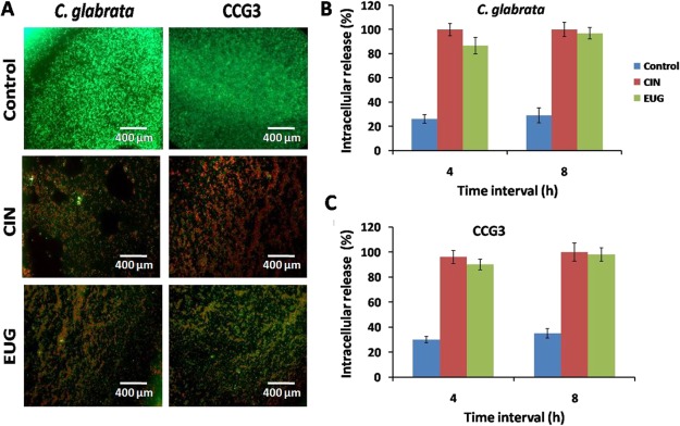
Figure 6.
(A) Fluorescence microscopy of 48 h mature C. glabrata and CCG3 biofilm after treatment with CIN (128 μg mL–1) and EUG (256 μg mL–1) stained with FDA + PI. Scale bar represents 400 μm. Quantification of intracellular material release in C. glabrata and CCG3 cells treated with (B) CIN (128 μg mL–1) and (C) EUG (256 μg mL–1).

