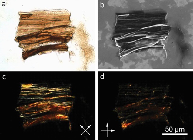
Figure 5.
(a) Optical and (b) corresponding SEM images of a piece of sediment obtained from slow addition of GO sheets into acetone. The sample was prepared by casting a droplet of the final dispersion containing the piece. In the SEM image (b), many flat sheets can be seen in the background that are neither crumpled nor collapsed. (c, d) Polarized optical microscopy images (polarizer directions marked by white arrows) indicate aligned microstructures along the horizontal wrinkles in this particle.

