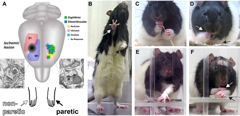
Figure 1.
(A) Ischemic lesions of the sensorimotor cortex result in impairments in the contralesional, paretic, forelimb (black arrows) and compensation with the nonparetic limb (white arrows). Synaptic structural plasticity in the contralesional motor cortex occurs due to denervation of transcallosal projections and increased reliance on the nonparetic limb. The residual cortex of the injured hemisphere can be driven to undergo greater neuronal plasticity with focused training (“rehabilitative training) of the paretic limb. A representative map of forelimb movement representations (green and blue, an average of n = 7 maps) is shown in the intact hemisphere. M1, primary motor cortex, S1, primary somatosensory cortex, *perforated synapse, scale bars = 400nm. (B) Rats rely more on the nonparetic limb for postural support. (C) In handling food pieces (in this example, a piece of uncooked vermicelli pasta), normal rats use dexterous forepaw movements. (D) After unilateral SMC lesions, rats use the paretic forelimb less and in less coordinated ways. (E) In skilled reaching, normal rats retrieve food pellets using a coordinated sequence of proximal and distal forelimb movements. (F) After unilateral SMC lesions, rats have movement abnormalities and frequently compensate with the nonparetic paw.

