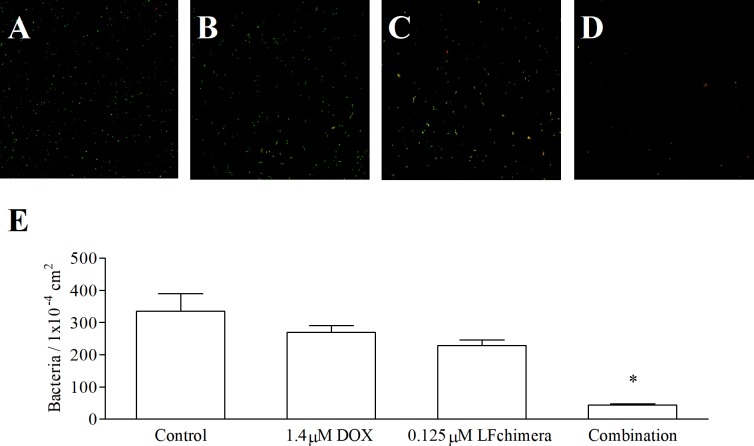
Fig 2. Effect of antimicrobial agent on adhesion state of biofilm-forming.
Confocal laser scanning micrographs of A. actinomycetemcomitans attached cells on glass coverslips after 30 min incubation in 1mM PPB (A), 1.4 μM DOX (B), 0.125 μM LFchimera (C) and combination of 1.4 μM DOX and 0.125 μM LFchimera (D). Bacteria were stained with LIVE/DEAD BacLight Bacterial Viability kit. Green color indicates live bacteria and red color indicates dead bacteria. Images were viewed at 630x magnification. (E) Bacterial count from 20 random areas of 2 coverslips for each sample. Data presented are the mean ± SD of adherent bacteria. *P < 0.01 compared with control and other agents.

