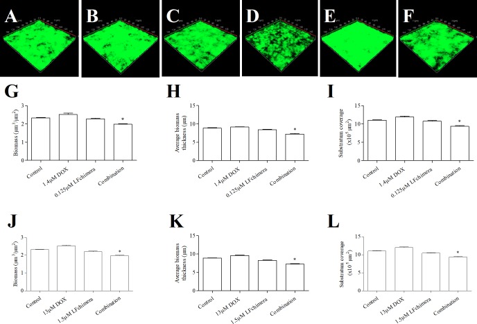
Fig 3. Effect of antimicrobial agent on biofilm formation.
CLSM 3-D reconstruction of 1-day old A. actinomycemcomitans ATCC 43718 biofilm after 15 min exposure to 1 mM PPB (A), 1.4 μM DOX (B), 0.125 μM LFchimera (C), combination of 1.4 μM DOX and 0.125 μM LFchimera (D), 13 μM DOX (E) and 1.5 μM LFchimera (F). The biofilms were stained with FITC-ConA. Green color indicates exopolysaccharide of biofilm matrix. CLSM-COMSTAT analysis comparing the effect of 1 mM PPB, 1.4 μM DOX, 0.125 μM LFchimera and the combination on biomass (G), average biomass thickness (H) and substratum coverage (I). CLSM-COMSTAT analysis comparing the effect of 1mM PPB, 13 μM DOX, 1.5 μM LFchimera and the combination on biomass (J), average biomass thickness (K) and substratum coverage (L). Values are means ± SD from 2 independent experiments. *P < 0.01 compared with control and other agents.

