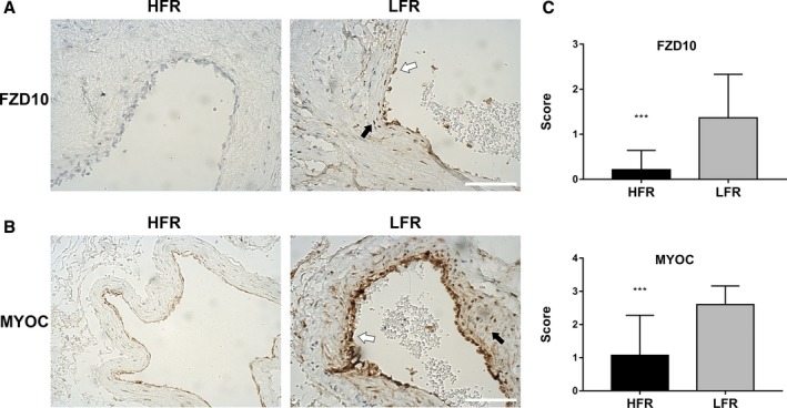
Figure 3.

Detection of FZD10 and MYOC in bAVM tissue samples. Immunohistochemical staining of bAVM tissue samples with differential flow rate subtypes show strong staining for FZD10 (A) and MYOC (B) in LFR bAVM tissue. Endothelial cells lining the vascular lumen (white arrows) and vascular smooth muscle cells in the vessel wall (black arrows) both show staining for FZD10 and MYOC. The scale bar corresponds to 200 μm. C, Semiquantitative grading of FZD10 and MYOC expression levels in the vascular structure of bAVMs. ***P<0.001. bAVM indicates brain arteriovenous malformations; HFR, high flow rate; LFR, low flow rate.
