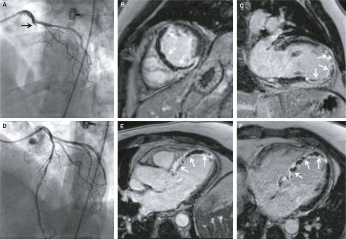
Figure 6.

Symptom‐to‐balloon time of 141 minutes: transmural necrosis and extensive MVO. A 48‐year‐old man presenting with chest pain and ST‐segment elevation in the anterior leads. Coronary angiography revealed TIMI 0 occlusion of the LAD (A); primary PCI with stent implantation was performed, resulting in TIMI 3 flow after PCI (D). Symptom‐to‐balloon time was 141 minutes. CMR revealed impaired left ventricular ejection fraction (38%) with akinesia in the anteroseptal, septal, and apical wall. Late gadolinium enhancement CMR images revealed extensive transmural necrosis with concomitant MVO (arrows) in the anteroseptal and septal wall (B, C, E) and the apex (E, F), consistent with the region supplied by the infarct‐related artery (large LAD). CMR indicates cardiac magnetic resonance; LAD, left anterior descending; MVO, microvascular obstruction; PCI, percutaneous coronary intervention; TIMI, Thrombolysis in Myocardial Infarction.
