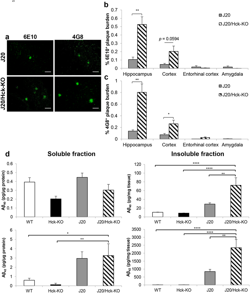
FIGURE 3.
Hck deficiency in J20 mice significantly increased Aβ plaque burden. (a) Representative images of 6E10- (left) and 4G8-positive (right) Aβ plaques found in the hippocampus region of 6–8 months old J20 (n = 8) and J20/Hck-KO (n = 6) mice. Scale bar, 50 μm. (b, c) Quantitative analysis of 6E10- (b) and 4G8-positive (c) Aβ plaques area found in the hippocampus, cortex, entorhinal cortex and amygdala regions of J20 and J20/Hck-KO mice. Significant accumulation of 6E10- and 4G8-positive plaques were observed in the hippocampus and cortex of J20/Hck-KO mice. Data are expressed as mean ± SEM from four sections per mouse. * p < 0.05 and ** p < 0.01 relative to J20 mice. (d) ELISA quantification of detergent-soluble (left) and -insoluble (right) Aβ40 (top) and Aβ42 (bottom) in protein lysates of mouse hippocampus. Lysates from WT, Hck-KO, J20 and J20/Hck-KO mice were analyzed at n = 6–8. Significant augmentation of insoluble Aβ40 and Aβ42 were detected when Hck was ablated in J20 mice. * p < 0.05, ** p < 0.01, and **** p < 0.0001 between indicated genotypes.

