FIGURE 5.
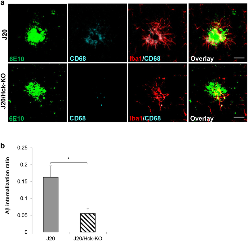
Knocking out Hck in J20 mice significantly reduced Aβ internalization in microglia. (a) Representative images of 6E10-positive plaques (green) clustered by Iba1-positive microglia (red) and CD68-positive microglial phagolysosomes (cyan) in J20 (top) and J20/Hck-KO (bottom) mice (6–8 months old). Scale bar, 20 μm. (b) Quantitative analysis of Aβ internalization ratio revealed a 66% reduction in microglial phagocytic activity in J20/Hck-KO mice. Aβ internalization ratio was calculated by volume of Aβ within microglial phagolysosomes normalized to microglia number on the plaque and Aβ plaque volume in the field. Plaques were analyzed in the hemibrains of 8 J20 (n = 18) and 6 J20/Hck-KO (n = 30) mice. Data are expressed as mean ± SEM from one section per mouse. * p < 0.05 relative to J20 mice.

