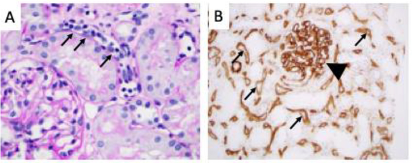
Figure 2. Histologic findings in antibody mediated rejection.
A) By light microscopy the peritubular capillaries are congested with leukocytes (“capillaritis”, small arrows). The adjacent tubules are intact and there is little infiltration of the tubules by leukocytes. B) Immunohistochemistry for C4d. C4d staining (brown) can be seen throughout the peritubular capillaries (small arrows) and within the glomerular capillaries (arrowhead).

