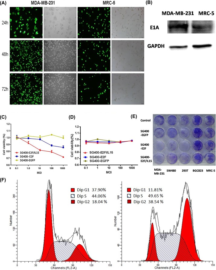
Figure 2. SG400-E2F/IL-15 selectively inhibited breast cancer cell proliferation.
(A) Representative photomicrographs were obtained from MDA-MB-231and MRC-5 infected with SG400-EGFP at the MOI of 1. Original magnification, 200×. (B) The expression of adenovirus structure protein E1A in MDA-MB-231 and MRC-5 cells 48 h after infection of SG400-E2F/IL15. 5 days after infection with SG400-E2F/IL15, MTT assay was performed to measure the proliferation of MDA-MB-231 (C) and MRC-5 (D) at different MOI (Panel C: SG400-EGFP and SG400-E2F, P=0.0002; SG400-E2F and SG400-E2F/IL15 P=0.0000. Panel D: SG400-EGFP and SG400-E2F, P=0.838; SG400-E2F and SG400-E2F/IL15, P=0.356). (E) Different cell lines (1 × 10⁵ cells/well) were cultured in 24-well plates and treated with SG400-EGFP, SG400-E2F, or SG400-E2F/IL15 (5 MOI) with PBS used as control. 3 days after infection, cytotoxicity was observed via crystal violet staining. (F) Cell cycle changed in MDA-MB-231 cells 5 days after infected with SG400-E2F/IL15 (10 MOI). Left panel: Uninfected MDA-MB-231 cells. Right panel: MDA-MB-231 cells infected with SG400-E2F/IL15.

