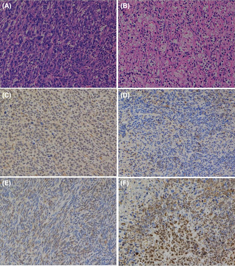
Figure 5. Pathological and immunohistochemical examination after infection with SG400-E2F/IL15.
HE staining of tumor tissues in PBS group (A) and SG400-E2F/IL15 group (B). Immunohistochemical analysis of E2F-1 (C,D) and IL-15 (E,F) of tumor tissues in PBS and SG400-E2F/IL15 group. Magnification 200×.

