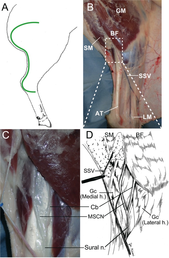
Figure 1.
Surgical procedure to identify of the ovine sural nerve. (A) The green line shows the incision line. From the lateral gluteal-thigh border to 2 cm posterior the lateral malleolus C-shape incision was made on the ovine cadaver right leg. (B) The gluteus maximus (GM), biceps femoris (BF), semimembranosus (SM), Achilles tendon (AT), lateral malleolus (LM) were dissected after undermining the skin flap. The short saphenous vein (SSV) was identified. (C,D) The sural nerve, medial sural cutaneous nerve (MSCN), and communicating branch (Cb) were identified to be located under the gastrocnemius fascia, between the lateral and medial head of the gastrocnemius, and distal of BF. Sural nerve formed by MSCN and Cb. Gc (Medial h.): Gastrocnemius medial head. Gc (Lateral h.): Gastrocnemius lateral head.

