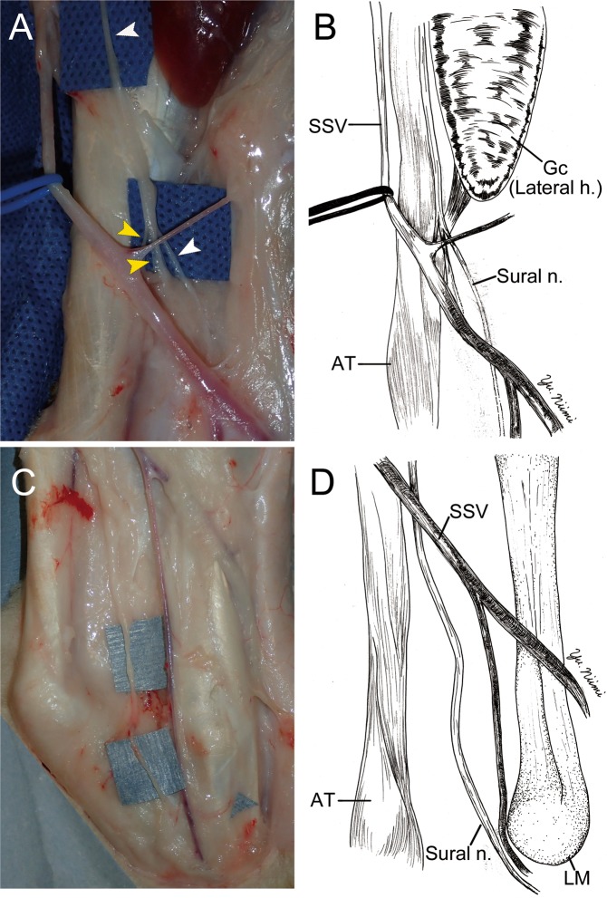
Figure 2.
The surgical anatomy of sural nerve (Sural n.); middle (A,B) and distal (C,D) portion of the leg. (A,B) The sural nerve (white arrow head) ran parallel to the short saphenous vein (SSV, blue rubber tape), and ran medial side of the gastrocnemius (Gc) lateral head (Lateral h.). Thereafter, two nerves (yellow arrow heads) were merging to the sural nerve at the midpoint of the lower leg. (C,D) In the distal portion of lower leg, the sural nerve was passed posterior to the lateral malleolus (LM). Thereafter, the sural nerve penetrated the deep fascia and went into the foot area. AT: Achilles tendon. Gc (Lateral h.): Gastrocnemius lateral head.

