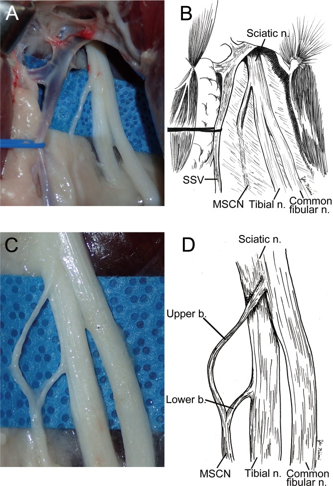
Figure 3.
The surgical anatomy of medial sural cutaneous nerve (MSCN). (A,B) To dissect to the proximal region from the point of Fig. 1, MSCN was divided from sciatic nerve (Sciatic n.) near the tibial nerve side between biceps femoris and semimembranosus. Common fibular nerve (Common fibular n.) was also devided from scaiatic nerve at the same place. (C,D) After removing connective tissue, MSCN was exposed forming from two branches, which were originated from sciatic and tibial nerves.

