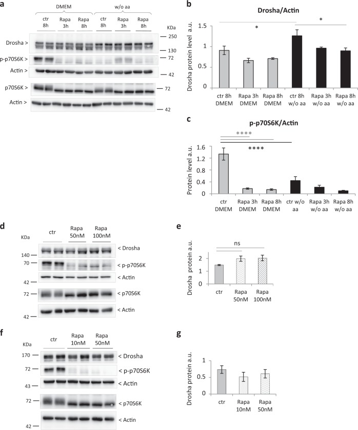
Fig. 5. Neither short- nor long-term rapamycin treatments are able to reproduce the increase of Drosha observed in aa-deprived primary adult rat hepatocytes.
a Western blot analysis of Drosha protein level, p70S6K total protein, and p70S6K phosphorylation. b Western blot quantification of Drosha protein in primary adult rat hepatocytes after 48 h aaD without any treatment (Rapa ctr), after 3 h and 8 h rapamycin treatment (100 nm) (Rapa 3 h, Rapa 8 h). Rapamycin was solubilized in DMSO, which was present in the rapamycin and control condition at a final concentration of 0.05 µl/mL. Values are means ± SEM (n = 3). *p < 0.05. c Western blot quantification of p70S6k phosphorylation in primary adult rat hepatocytes after 48 h aaD without any treatment (Rapa ctr), after 3 h and 8 h rapamycin treatment (100 nm) (Rapa 3 h, Rapa 8 h). Note that p70S6K phosphorylation was measured as read out of rapamycin effect on mTOR activation. Values are means ± SEM (n = 3). ****p < 0.0001. d Western blot analysis of Drosha protein level, p70S6K total protein and p70S6K phosphorylation and e western blot quantification of Drosha protein in aa-deprived hepatocytes without any treatment (ctr) and rapamycin treatment for 24 h. Values are means ± SEM (n = 3). f Western blot analysis of Drosha protein level, p70S6K total protein, and p70S6K phosphorylation and f western blot quantification of Drosha protein in aa-deprived hepatocytes without any treatment (ctr) and rapamycin treatment for 48 h. Values are means ± SEM (n = 5)

