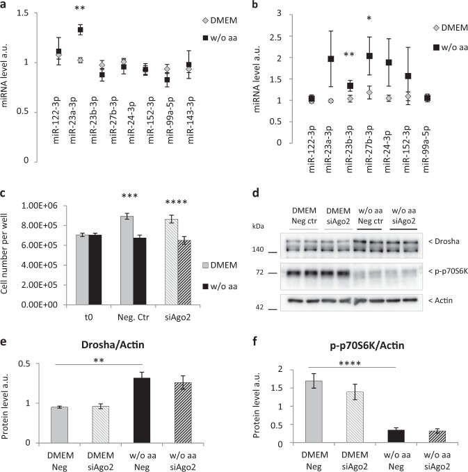
Fig. 8. AaD in primary adult rat hepatocytes increases the expression of miR-23a/b and miR-27b, but Ago2 knockdown has no effect either on cell proliferation or on Drosha protein.
Analysis of miRNA expression level in primary adult rat hepatocytes after 24 h a and 48 h b either in full-aa medium (DMEM) or medium w/o aa. RNA was extracted, reverse transcribed and analyzed by Real Time PCR. The expression of miR-23a-3p, -23b-3p, -27b-3p, -24-3p, -152-3p, -99a-5p, -143-3p was normalized to the miR-122-3p transcript level, whereas the expression of miR-122-3p was normalized to miR-99a-5p. Values are means ± SEM (n = 3). *p < 0.05, **p < 0.005. c Cell counting of primary adult rat hepatocytes transfected with 50 nmol/L negative control-siRNA (Neg. Ctr) or Ago2-siRNA (siAgo2) for 48 h in full-aa medium (DMEM) or in absence of aa (w/o aa). Values are means ± SEM (n = 3). ***p < 0.0001, ****p < 0.00001. d Western blot analysis of Drosha protein level and p70S6K phosphorylation in primary adult rat hepatocytes transfected with 50 nmol/L negative control-siRNA (Neg. Ctr) or Ago2-siRNA (siAgo2) for 48 h in full-aa medium (DMEM) or in absence of aa (w/o aa); e, f western blot quantification of Drosha protein e and p70S6K phosphorylation f in aa-deprived hepatocytes transfected with Ago2-siRNA or the negative control, compared with hepatocytes in full-aa medium. Values are means ± SEM (n = 3). **p < 0.001, ****p < 0.00001

