
Figure 4.
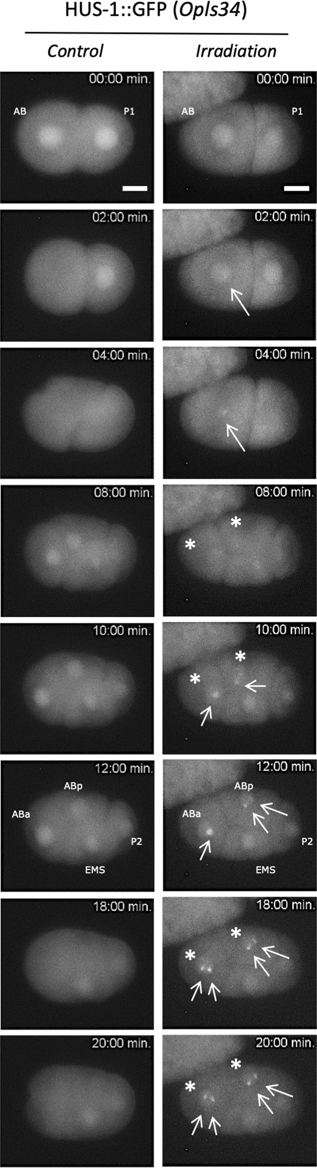
Real-time analysis of micro-irradiated AB nucleus revealed the subcellular relocalization of HUS-1::GFP (foci). First cell divisions of 2-cell stage embryos are observed using a strain expressing HUS-1::GFP (opls34). Before irradiation HUS-1::GFP is homogenously distributed in nuclei (t = 0 min). The AB cell nucleus is targeted with protons at t = 0 min. In micro-irradiated AB nucleus of HUS-1::GFP embryo, a focus, indicated with white arrow (→), appears just before the first cell division of AB (t = 2 min.) and reappears in the daughter cells ABa and ABp indicated with stars (*, right). We never observed foci in both non-irradiated embryo (control) and neighbouring non-irradiated nuclei (P1, EMS, and P2). Irradiated embryos (opls34 = 104 protons) were observed in real time following irradiation. Scale bar: 10 µm.