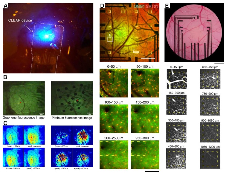
Figure 2.
Integration of transparent electrodes with complementary optical techniques in vivo. (A) Gr-based electrodes implanted on a mouse cerebral cortex. An optical fiber is also shown and used to deliver optogenetic stimulation. Modified with permission from Park et al. (2014). (B) Gr-based electrodes implanted on a mouse cerebral cortex (left) or traditional platinum electrodes Howe et al. (2013) and peak calcium responses from the same regions (C). Panels (B,C) are from Park et al. (2018). Copyright ACS Publication. (D) Two-photon calcium imaging to a depth of 300 μm through Gr-based electrodes and intravascular imaging (E) to a depth of 1,200 μm. Modified with permission from Thunemann et al. (2018).

