
Figure 4.
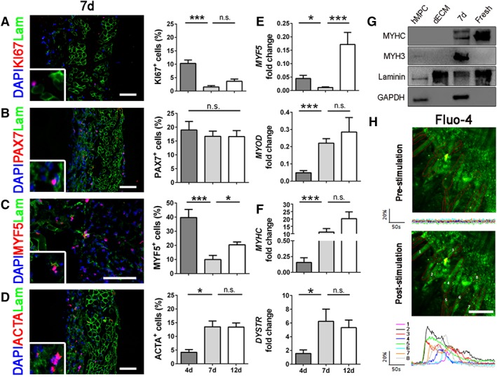
Immunofluorescence, quantifications, and molecular biology analyses of early and late myogenic markers. (A): Representative immunofluorescence images of recellularized samples after 7 days. KI67 (red), laminin (green), and nuclei counterstained with DAPI (blue). Quantifications were done at days 4, 7, and 12. (B): Immunofluorescence of satellite cells marker PAX7 (red) and related quantifications. (C): Immunofluorescence and quantifications of the early myogenic marker MYF5 (red). (D): Immunofluorescence and quantification of the late myogenic marker ACTA (red). (E): Molecular biology analyses (real‐time PCR) at 4, 7, and 12 days of early myogenic markers MYF5 and MYOD. (F): Molecular biology analyses of late myogenic markers MYHC and DYSTR. (G): Western blot analyses of mature myogenic proteins. GAPDH was used as human specific cell marker, laminin as extracellular matrix control. (H): Representative images of recellularized samples after 7 days in culture with myotubes loaded with the calcium dye Fluo‐4 AM and stimulated by ATP application. Graphics show mean fluorescence intensity (ΔF/F0) of the Region of Interests depicted. All scale bars: 100 μm. *, p < .05; **, p < .01; ***, p < .001; n.s., not significant by Mann–Whitney U test.