
Figure 3.
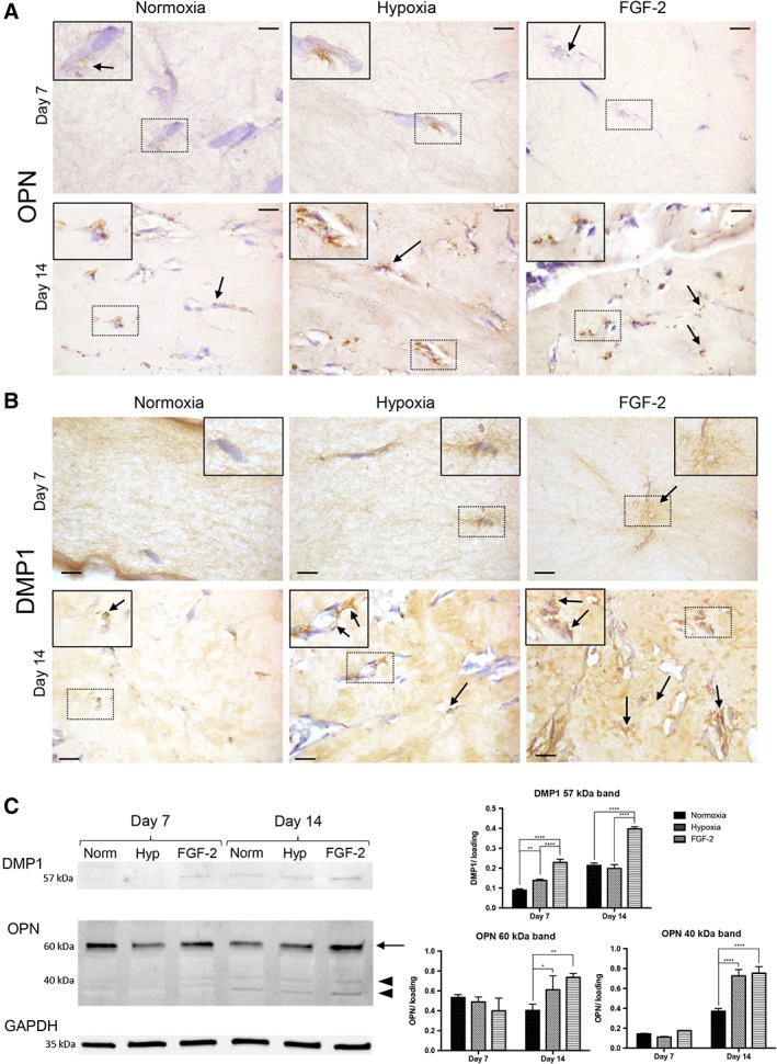
Osteogenic differentiation of stem cells from human exfoliated deciduous teeth in tissue‐engineered scaffolds. (A, B): Representative images of osteopontin (OPN; A) and dentin matrix protein 1 (DMP1; B) immunohistochemistry showing, for all the conditions, positive immunolabeling (arrows) adjacent to the cells at day 7 and day 14. Scale bars: 10 μm. Inset detail showing the area of interest at higher magnification. (C): Western blotting of DMP1 and OPN with a normalized quantitative analysis. DMP1 expression showed an increased abundance of the 57 kDa fragment in the FGF‐2 condition (×2.5 and ×1.8 at days 7 and 14, respectively) compared with the hypoxia and control groups. At day 14, OPN showed an increased abundance of 40 kDa (arrowhead) and 60 kDa (arrow) forms in the primed groups, especially FGF‐2 group (×1.9) compared with the control group. No significant difference was observed at day 7 for both OPN forms. Each band was normalized using GAPDH band as housekeeping protein (n = 3 per group). *, p < .05; **, p < .01; ****, p < .0001.