
Figure 5.
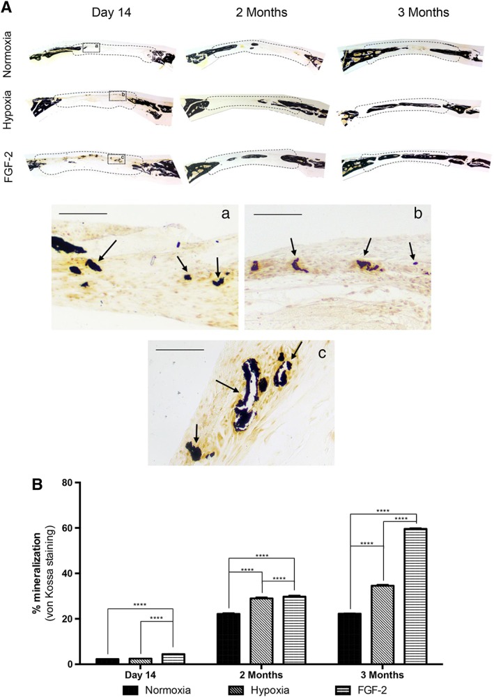
Mineral formation in calvarial bone defects. (A): von Kossa staining revealing bone formation (arrows) in all the conditions within the defects (delineated by a dotted line), at day 14, and 2 and 3 months. Inset detail at day 14: normoxia (Aa), hypoxic priming (Ab), and FGF‐2 priming (Ac). Scale bars: 50 μm. (B): Quantitative analysis of von Kossa staining showing that mineral formation was significantly increased in FGF‐2 primed samples at all‐time points when compared with control (stem cells from human exfoliated deciduous teeth with no priming) or hypoxic primed samples. At 2 and 3 months, bone formation was also increased in the defects filled with hypoxia primed constructs when compared with control constructs. ****, p < .0001.