
Figure 7.
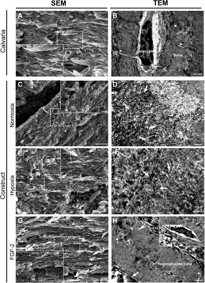
Ultrastructural analysis of the grafted calvaria at 3 months postimplantation. (A, C, E, G): Representative SEM image for each condition revealing the different areas of the calvaria: grafted and original part (scale bar: 5 μm). Inset detail showing collagen fibers organization within the defect and the calvaria at higher magnification (scale bar: 1 μm). (B, D, F, H): Representative transmission electron microscopy image for each condition revealing the ultrastructural difference for each area and conditions (scale bar: 1 μm). (A, B): Representative image of the native mouse calvarial bone showing well‐organized collagen fibers with similar same orientation. Osteocytes were observed in their lacuna associated with their delicate network of canaliculi (asterisks). (C, D): Heterogeneous matrix and poorly organized collagen fibers (white arrows) were observed in the control (normoxia) constructs at both magnifications. (E, F): In hypoxia primed constructs, the collagen fibers (white arrows) were more packed and better organized. Lacuno‐canalicular system (asterisks) was observed in the constructs. (H, I): In FGF‐2 primed constructs, the fibers (white arrows) were well‐aligned and showed a dense organization very similar to the adjacent bone. Of note, well‐distinguishable osteocytes (inset) with their lacuno‐canalicular system (asterisks) were observed associated with dense regenerated bone matrix in the construct.