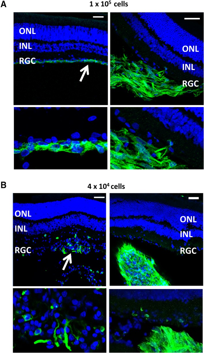
Figure 6.

Localization of transplanted cells following immunostaining or retinal sections with antibodies to both human nestin plus CD29 and Alexa flour 488 used as a single secondary antibody. Images show eyes transplanted with (A) 1 × 10⁵ cells and (B) 4 × 10⁴ cells. White arrows indicate the location of the cells. Scale = 50 μm. Abbreviations: INL, inner nuclear layer; ONL, outer nuclear layer; RGC, retinal ganglion cell layer.
