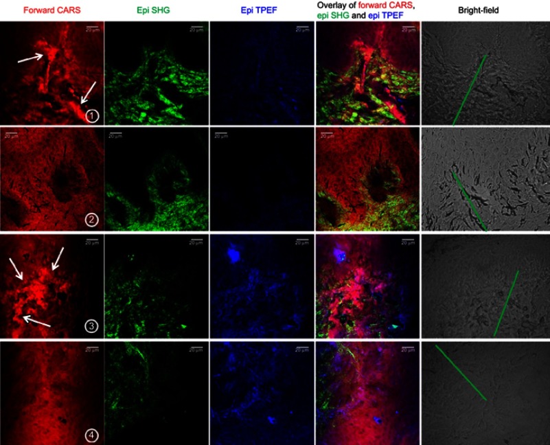
Figure 3.
Non-linear microscopy of dermal region of skin tissue for healthy and treated biopsies: display of forward CARS, epi SHG, and epi TPEF, overlay of CARS, SHG, and TPEF, and bright-field images in the dermis region of the skin for each sample condition. Row 1: lesional psoriasis tissue before treatment; row 2: psoriasis tissue treated with NBUVB; row 3: healthy tissue; and row 4: healthy tissue treated with NBUVB. The bright-field images indicate the dermis (green line). Scale bar: 20 µm.
Abbreviations: CARS, coherent anti-Stokes Raman spectroscopy; NBUVB, narrowband ultraviolet B; SHG, second-harmonic generation microscopy; TPEF, two-photon excitation fluorescence microscopy.

