Fig. 3.
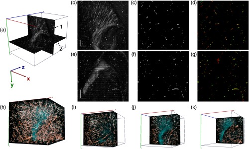
Deep-learning-based vessel segmentation at OCT natural resolution. (a) Sagittal (1) and coronal (2) planes of 3-D dataset. (b) Sagittal and (e) coronal views of cross-polarization images. Vessel segmentation from (c) the sagittal and (f) coronal images. Composite images in (d) and (g) show two separate segmentation results from sagittal (red) and coronal (green) views. (h) 3-D reconstruction of vasculature in a brain volume of . In addition, cross-polarization intensity is given in cyan. (i)–(k) Three composite thin layers () of the same dataset. The -stack of the whole volume is shown in Video 1 (MP4, 11.1 MB [URL: https://doi.org/10.1117/1.NPh.6.3.035004.1]). Scale bar: .

