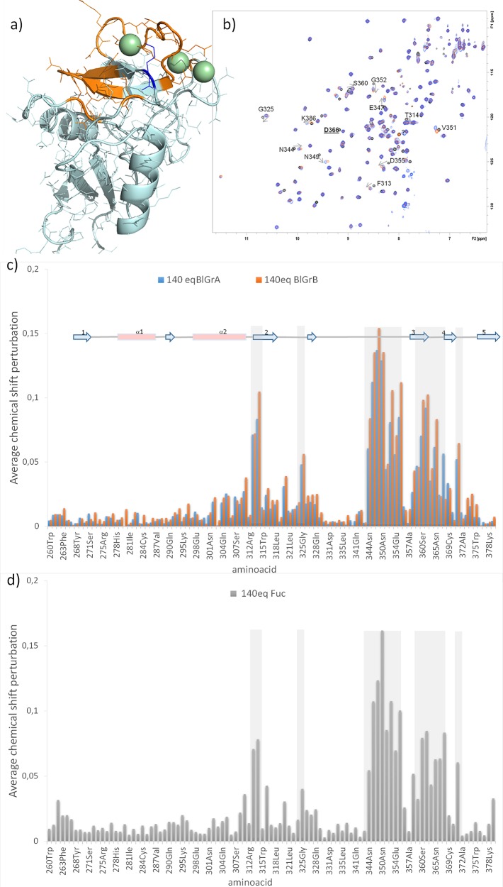
Figure 1.
Chemical shift perturbations for the interaction of CRD DC-SIGN with tetrasaccharides (1 and 2) and with Fuc (4). (a) In orange amino acids with chemical shifts most perturbed upon the addition of BGB and BGA. In blue K368, affected more with BGA than with BGB. (b) Superimposition of 1H–15N HSQC spectra (black, apo DC-SIGN; orange, in the presence of 140 equiv of BGB; blue, in the presence of 140 equiv of BGA). Some affected crosspeaks are annotated. Residue D366 that disappear in the middle points of the titration is underlined. (c, d) Average chemical shift perturbation upon the addition of BGA, BGB and Fuc. [D366 is not included in the plot; average chemical shift perturbations were calculated using the formula {1/2[δH2 + (0.2δN)2]}1/2, where δH and δN are the chemical shift change in 1H and 15N, respectively (in ppm), between the apo and bound forms.]

