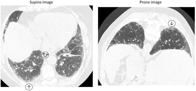
Figure 3: HRCT in SSc-ILD.

Inspiratory and prone HRCT demonstrating minimal reticulation, subpleural groundglass opacity (↑) that persists on prone imaging (↓) suggestive of interstitial lung abnormalities and an early fibrotic lung disease. No honeycombing or traction bronchiectasis. Note slightly dilated esophagus (*).
