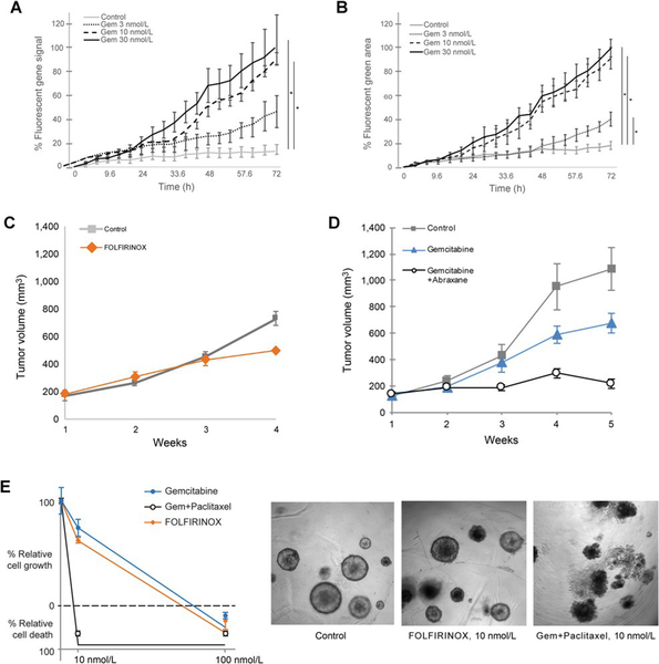
Figure 6.
Patient-derived models respond differentially to chemotherapeutic agents. A and B, Real-time levels of apoptosis for PDX-derived organoids from patient 1 (A) and tumor-derived organoids from patient 6 (B) were treated with gemcitabine (3 nmol/L, 10 nmol/L, 30 nmol/L). Apoptosis was measured in real-time over 72 hours (% fluorescent green signal, n ¼ 4). PDX from patient 1 (as in A), was treated in vivo with FOLFIRINOX (10 nmol/L; C), or gemcitabine (10 nmol/L) with or without abraxane (10 nmol/L; D). Tumors growth (mm3) was measured over 4 weeks. Control mice received vehicle, (n ¼ 3). Points are mean tumor volume; bars, SE. (C and D). The same treatment was applied in vitro to PDX-derived organoids from patient 1. Proliferation was measured by % relatively cell growth/death (E), after 72 hours of growth. SE, *, P < 0.001. Representative light microscopy (10x) images at 10 nmol/L are shown (right).

