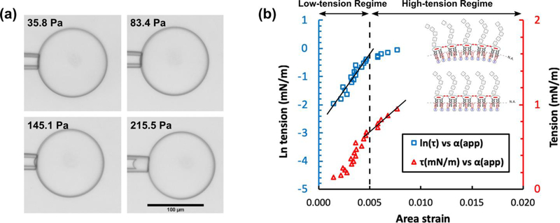
Fig. 5.
The micropipette aspiration method tested on GUVs possessing DOPC/DPPC/Chol (3:3:4 mol%) and LPS in the inner and outer leaflets, respectively. (a) As is depicted in the four panels, by increasing the withdrawing hydrostatic pressure (i.e. from 36 to 215 Pa) a portion of the lipid bilayer membrane was drawn into the micropipette aspirator. The scale bar denotes 100 μm and is the same in all panels. (b) The plot shows the logarithm of tension (i.e. blue squares) and tension (i.e. red triangles) versus the apparent area strain. The lines are fitted to the blue squares and red triangles in the low- and high-tension regimes, respectively. The R-squared values are larger than 90%. The schematics in the inset illustrate our hypothesis on the effect of repulsive electrostatic stress between the lipid A section of LPS molecules on the bending and area expansion moduli (see text).

