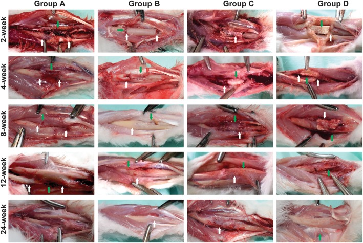
Fig. 2.
Gross observation post-implantation. Group a at the 2nd week: the bone defect was obvious (white arrow), massive tissue fluid exudation, and formation of fresh bone tissues (green arrow); there are no signs of infection. Group b at the 2nd week: the bone stump neat (white arrow); massive fibrous tissue formed in the gap (green arrow). Group c at the 2nd week: the boundary between the BCBB and the bone stump was clear, with fibrous tissue filling and no obvious signs of infection (white arrow). Group d at 2nd week: there was no significant difference between group d and group c, the structure of the material was complete, the gap between the host and the material was filled with fibrous tissue (white arrow), there was new tissue ingrowth in the pores of the material (green arrow), and no obvious signs of infection. Group a at the 4th week: the exudation was reduced and the medullary cavity was closed by fibrous tissue (white arrow) and fibrous tissue adhesion (green arrow). Group b at the 4th week: at the bone stump, formed the fibrous callus (white arrow) and periosteal tissue formed on the surface of the autologous bone (green arrow). Group c at the 4th week: the surface of the material was complete, the host bone tissue is closely connected (white arrow), and the callus wrapped around the surface of the material (green arrow). Group d at the 4th week: the surface structure of the material was complete, and the material was closely connected with the host bone (white arrow), surrounded by the callus, rich in blood supply (green arrow). Group a at the 8th week: the medullary cavity was closed by the bone tissue and surrounded with a large amount of fibrous tissue and muscles (white arrow), and the exudation was reduced and absorbed (green arrow). Group b at the 8th week: the callus becomes bony union (white arrow), and the growth of the callus and the resorption and reconstruction of the bone graft were obvious (green arrow). Group c at the 8th week: the material contour was complete, and the BCBB was closely connected to the host bone (green arrow), the surface of the material and the pores were filled with callus, and the material was absorbed and degraded in different degrees (white arrow). Group d at the 8th week: the material structure is not as complete as 4 weeks, the surface can be seen a large number of callus formation (white arrow), the material and the host bone tightly connected, and the material is absorbed; repair section had a good shape (green arrow). Group a at the 12th week: bone resorption in the area of the bone defect (black arrow), the surrounding fibrous tissue formed fibrous scar (green arrow). Group b at the 12th week: the autogenous bone was completely fused with the original bone (white arrow), the surface was covered with periosteum-like tissue, and the blood supply was abundant (green arrow). Group c at the 12th week: materials and bone tissue boundary are not clear (green arrow), and the repair section had a good shape (white arrow). Group d at the 12th week: the material and the bone formation are firm, the boundary is not clear, the material was partially absorbed, the new bone has grown, and the partial shape approaches the normal radius (green arrow). Group a at the 24th week: there was no significant difference between 12 and 24 weeks of observation. Group b at the 24th week has the same shape close to the radius in 24 weeks (white arrow) and 12 weeks. Group c at the 24th week: the surface of the material is rich with callus, rich in blood supply, and the boundary between the host bone tissue is not clear and the repair section had a better shape than 12 weeks (black arrow). Group d at the 24th week: the repair section was well shaped, and there was no significant difference between the host bone and material (green arrow)

