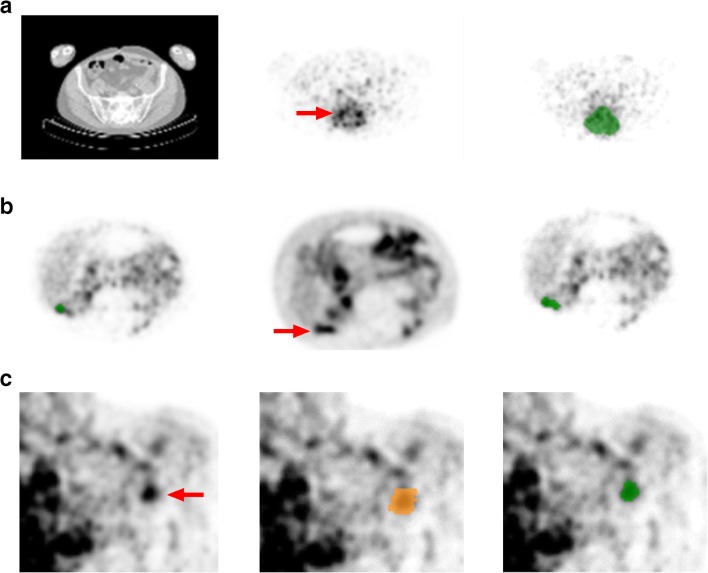
Fig. 2.
VOI delineation methods for 89Zr-immuno-PET. Manual tumor delineation on immuno-PET (a) using the low dose CT (left panel), attenuation corrected 89Zr-cetuximab-PET on D6 (middle panel) with tumor lesion indicated by the red arrow and example VOI on 89Zr-cetuximab-PET shown in green (right panel). Manual tumor delineation on immuno-PET after reviewing the corresponding 18F-FDG-PET (b) the original manually delineated VOI shown in green on the 89Zr-trastuzumab-PET on D4 (left panel), reviewing the 18F-FDG-PET scan with tumor lesion indicated by the red arrow (middle panel) and adapting the original VOI after reviewing the 18F-FDG-PET scan; the FDG adapted tumor VOI shown on 89Zr-trastuzumab-PET is in green (right panel). Semi-automatic delineation (c) with the attenuation corrected 89Zr-rituximab-PET on D6 (left panel), the mask delineated on the 89Zr-rituximab-PET shown in orange (middle panel) and the semi-automatic VOI (50% of ACpeak, mask restricted) on the 89Zr-rituximab-PET shown in green (right panel). This semi-automatic VOI was accepted by the observer, as it contains tumor and no other structures or background

