
Fig. 1.
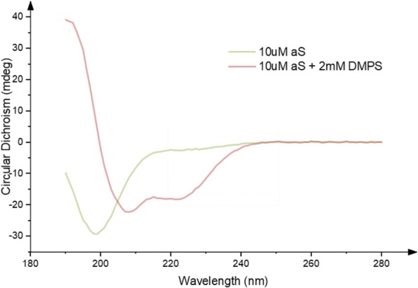
Change in Circular Dichroism (CD) signal in the far UV caused by the binding of αS to an excess of DMPS vesicles. This demonstrates a shift from a random coil structure in the absence of lipid vesicles (green), towards an alpha-helical secondary structure in the presence of DMPS lipid vesicles (red). Meade et. al. unpublished data reproducing data from Galvagnion et al. [34]