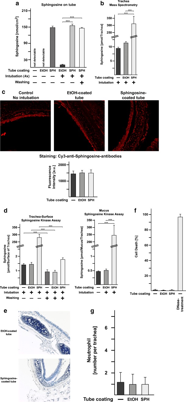
Fig. 6.
Sphingosine coating of endotracheal tubes is stable in vivo, non-toxic to tracheal epithelial cells, and does not induce inflammation. a Tubes were sphingosine (SPH) or ethanol (EtOH)-coated and inserted over a total of 4 h into mice. To avoid variations due to prolonged and very deep anesthesia, we intubated 4 mice consecutively on 4 days for 1 h each. The concentration of sphingosine on the tube was determined by a sphingosine-kinase assay. Sphingosine concentrations on the tubes remained stable and even slightly increased, very likely by binding sphingosine-containing mucus, which is removed by washing the tube. Likewise, ethanol-coated tubes contained low amounts of sphingosine on the surface after intubation. Ethanol-coated or uncoated tubes (not used for intubation) were negative. Shown are the mean ± SD of sphingosine concentrations in pmol/cm2, n = 6 each, ANOVA, *p < 0.05, **p < 0.01, ***p < 0.001. b Mass spectrometry reveals that intubation of sphingosine-coated tubes results in a marked increase of total sphingosine in the trachea. Mice were intubated as above for 60 min, the tube removed, the trachea removed and subjected to mass spectrometry. Shown is the mean of the concentration of sphingosine ± SD, n = 3–5 each, ANOVA, *p < 0.05, **p < 0.01, ***p < 0.001. c Immunofluorescence microscopy analysis of paraffin sections of trachea stained with Cy3-coupled anti-sphingosine antibodies does not reveal an increase of sphingosine in the epithelial cells of the trachea or the submucosa or any other cell of the trachea. Shown are typical results and the mean ± SD of the quantification of the fluorescence staining of each 100 cells of the trachea from 6 independent samples per group. The fluorescence intensity is given in arbitrary units. d In situ surface sphingosine-kinase assays on trachea from intubated mice reveal that most of the sphingosine released from the tube remains in the mucus of the trachea. Washing the trachea prior to the kinase assay removes the increase of sphingosine detected after intubation, which is recovered in the mucus in the wash buffer. Shown is the mean of the concentration of sphingosine ± SD per trachea, n = 6 each, ANOVA, *p < 0.05, **p < 0.01, ***p < 0.001. To exclude toxic or pro-inflammatory effects of sphingosine-coated tubes, we performed hemalaun, Cy3-coupled anti-Gr1-antibody and TUNEL stainings of paraffin sections of the trachea from mice that were intubated with sphingosine- or ethanol (EtOH)-coated ventilation tubes. We did not observe any e structural damage, f induction of cell death, or g influx of granulocytes/monocytes into the submucosa epithelial cell layer in the trachea of mice intubated with sphingosine-coated plastic tubes. Displayed are typical examples of the stainings and the quantitative analysis of the number of dead epithelial cells and the influx of leukocytes, which were quantified in 10 sections from each trachea from 6 mice, i.e., a total of 60 sections. In each section, the entire epithelial cell layer and the submucosa of the trachea were investigated. Structural damage was determined as the number of disruptions of the epithelial cell layer. Shown are the mean ± SD, n = 6 each, ANOVA, *p < 0.05, **p < 0.01, ***p < 0.001

