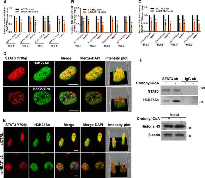
Fig. 8.
NEAT1 influences STAT3 binding to endocytosis-related genes. The siNEAT1 and siCTRL cells were collected for ChIP assays to analyse the relative fold enrichment of the CAV2 (a), TGFB2 (b), or TGFBR1 promoter (c) with anti-STAT3 or RNAP II antibodies. The data points represent the mean values determined from three independent experiments. The data are presented as the mean ± SD. d The U251 cells were fixed and incubated with anti-STAT3 (red), anti-H3K27Ac (green) or anti-H3K27Cro antibodies (green) before the confocal analysis. The intensity plots for the red and green channels were analysed with the ImageJ software. Scale bars 10 μm. e After transfection with the siNEAT1v2 or negative control siRNA, the U251 cells were fixed and incubated with anti-STAT3 (red) or anti-H3K27Ac antibodies (green) before the confocal analysis. The intensity plots for the red and green channels were analysed with the ImageJ software. Scale bars 10 μm. f After 24 h of incubation with 80 µm crotonyl-CoA or the mock control, the U251 cell lysates were harvested and subjected to an immunoprecipitation assay with anti-STAT3 or anti-IgG antibodies. The retrieval of STAT3, H3K27Ac, H3K27Cro+ and Histone H3 by endogenous STAT3 and IgG was measured by western blotting. *p < 0.001

