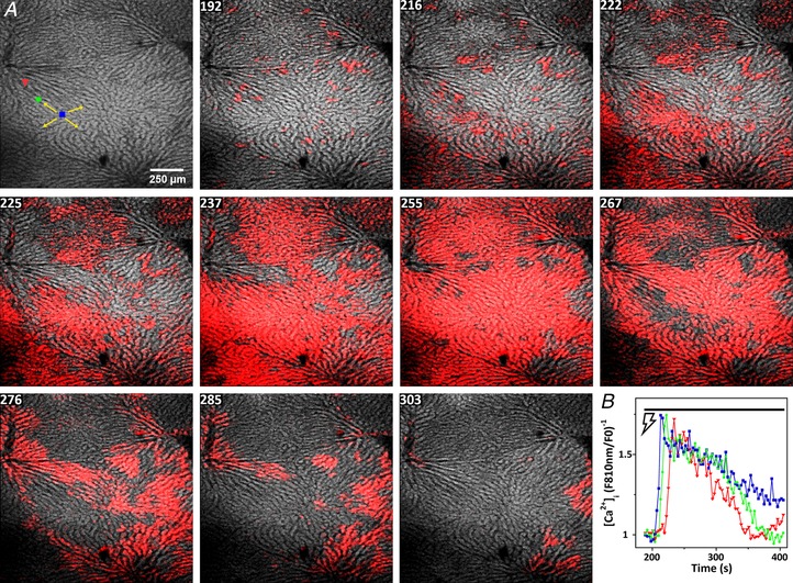
Figure 6. Electrical stimulation synergizes with the Ca2+‐responses induced by low concentrations of vasopressin.

Fura‐2/AM loaded liver was perfused with a subthreshold dose of vasopressin (10 pm, panels 150–192 s) prior to stimulating the hepatic nerve fibres at 4 V and 4 Hz starting at 195 s. Time is indicated (s). The figure corresponds to Movie S5 in the Supporting information. A, blue, green and red ROIs represent three hepatocytes along a hepatic plate from the periportal to pericentral regions, respectively. B, time courses of the electrically‐induced [Ca2+]c increases in the ROIs.
