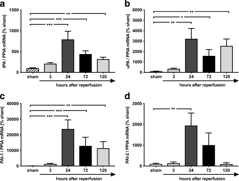
Fig. 2.
PAI-1 and PAI-2 is strongly regulated after MCAO. In tissue of ischemic injury, the expression of tPA and uPA peaked at 24 h (tPA ***p < 0.001 vs. sham (a); **uPA p < 0.01 vs. sham (b)). Their opponents, PAI-1 and PAI-2, were massively upregulated after MCAO. c PAI-1 showed an increase at 3 h with a peak expression at 24 h (237-fold increase, ***p < 0.001 vs. sham). d Expression of PAI-2 showed a 19-fold increase at 24 h post insult (**p < 0.01 vs. sham). Data are shown as mean ± SEM (n = 6 per group; sham: n = 2 per each group). Descriptive p values are assigned as follows: *p < 0.05, **p < 0.01, ***p < 0.001

