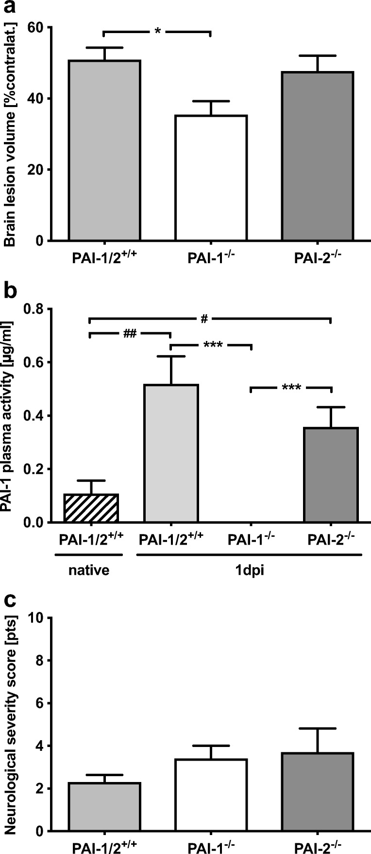
Fig. 4.

Reduced infarct volume in PAI-1-deficient mice. a The infarct volumes were reduced in PAI-1-deficient mice by 31% compared to wild-type mice 24 h after insult (PAI-1−/− 35 ± 11% of contralateral hemisphere, *p < 0.05; PAI-1+/+/PAI-2+/+ 51 ± 10% of contralateral hemisphere). PAI-2 deficiency did not influence brain injury (48 ± 13% of contralateral hemisphere). b The PAI-1 plasma activity was undetectable in PAI-1-deficient mice but increased over time without differences between wild-type or PAI-2-deficient mice (PAI-1+/+/PAI-2+/+ 5-fold increase, ##p < 0.01 vs. native; PAI-2−/− 3.5-fold increase, #p < 0.05 vs. native). c The neurological severity score showed no differences between groups. Data are shown as mean ± SEM (n = 10 per group; native n = 4). Descriptive p values are assigned as follows: *p < 0.05, **p < 0.01, ***p < 0.001 using the Kruskal-Wallis test; #p < 0.05, ##p < 0.01, ###p < 0.001 using Welch’s test
