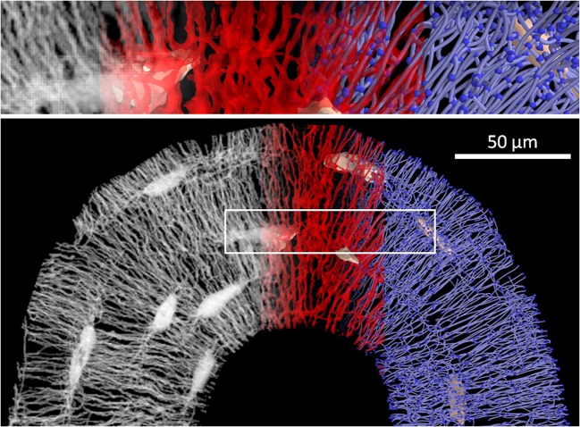
Fig. 1.
Below: work flow from an image stack obtained by confocal laser scanning microscopy (CLSM) (gray, left) to a binarized image of the LCN (red, middle) to a mathematical network consisting of edges (i.e., canaliculi) and nodes (i.e., lacunae and meeting points of canaliculi) (blue, right). The image at the top shows a magnification of the volume encircled by the white box

