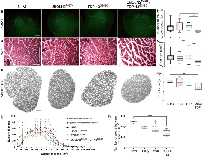
Fig. 4.
Motor neuron loss, axonal degeneration, and muscle atrophy. a Immunofluorescence of ChAT in lumbar ventral horn at 8 months of age. ChAT staining is specific to motor neuron. Pictures were taken at × 20 magnification (scale bar = 100 μm). b Number of motor neuron (MN) per ventral horn (three mice, six sections per mice). c Hematoxylin & eosin (H&E) coloration of transverse section of gastrocnemius muscle at 8 months of age. Pictures were taken at × 10 magnification with light microscopy (scale bar = 200 μm). d Muscle fiber size in μm2 (three mice, three sections per mice. e Epoxy-fixed ventral root innervating gastrocnemius muscle at 8 months of age. Pictures were taken at × 20 magnification with light microscopy (scale bar = 100 μm). f Total ventral root area in μm2 (three mice, three sections per mice). Area were all manually measured with ImageJ software. g Number of axons per caliber interval in μm2. Only significant results between double transgenic and NTG (¥, p ˂ 0.05) and between double transgenic and TDP-43G348C are shown (‡, p ˂ 0.05). Results are also significant between double transgenic and UBQLN2P497H (not illustrated). h Number of large axons between 35 and 80 μm2 combined in one interval

