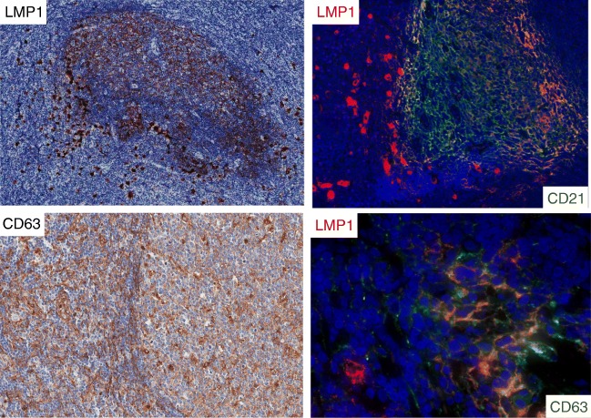
Fig. 4.
Case 1. Numerous LMP1+ large cells surround and partially infiltrate B cell follicles with LMP1+ FDCs. Double staining with immunofluorescence confirmed the presence of peri-follicular EBV-infected cells (LMP1+, red) and of CD21+ reticular FDCs (green). CD63, a marker for exosomes, was present in most stromal cells with reticular morphology inside and outside GCs. A high magnification of GC revealed the presence of double-positive cells (LMP1/CD63) with dendritic morphology

