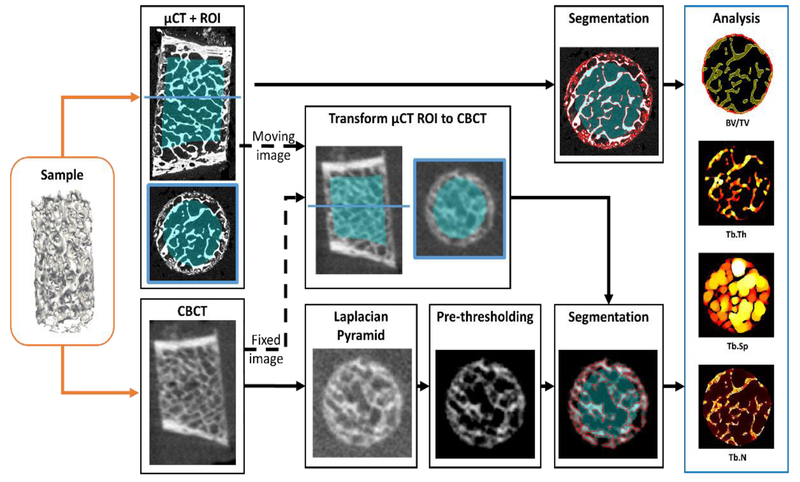
Figure 2.
Workflow of the evaluation study. Each bone sample was imaged on extremity CBCT and gold standard μCT. A binary trabecular mask was created in the μCT volume and transformed to the CBCT volume to generate ROIs for analysis of metrics of trabecular microstructure. To enable robust trabecular measurements in CBCT, segmentation algorithms ranging from global Otsu’s thresholding to local thresholding with Laplacian Pyramid image enhancement were evaluated.

