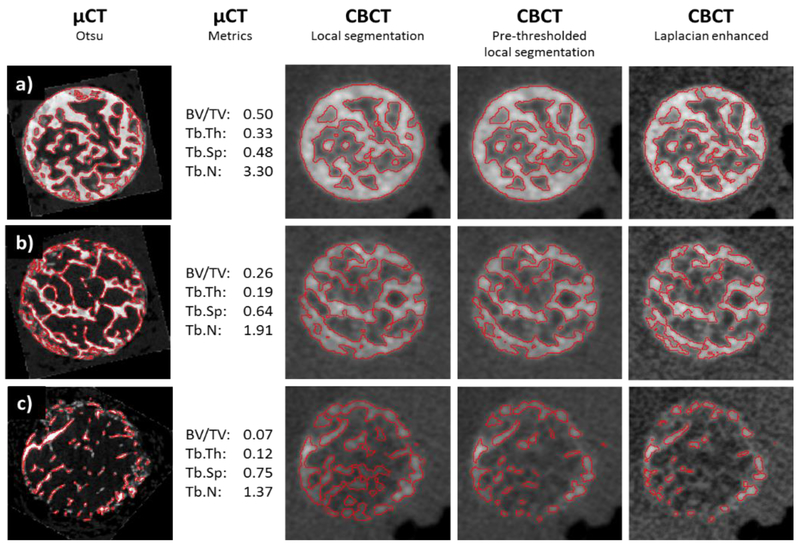
Figure 4.
Comparison of three samples (a, b and c) imaged on μCT and CBCT and processed with different pre-processing and segmentation methods. First column shows axial view of μCT images with an overlay of a global Otsu’s segmentation. Values of trabecular metrics are also provided. The subsequent columns show the same sample imaged on extremity CBCT and segmented using local thresholding, local thresholding with a global pre-thresholding step, and Laplacian-enhanced local thresholding. To facilitate visual comparison, μCT images were registered to CBCT.

