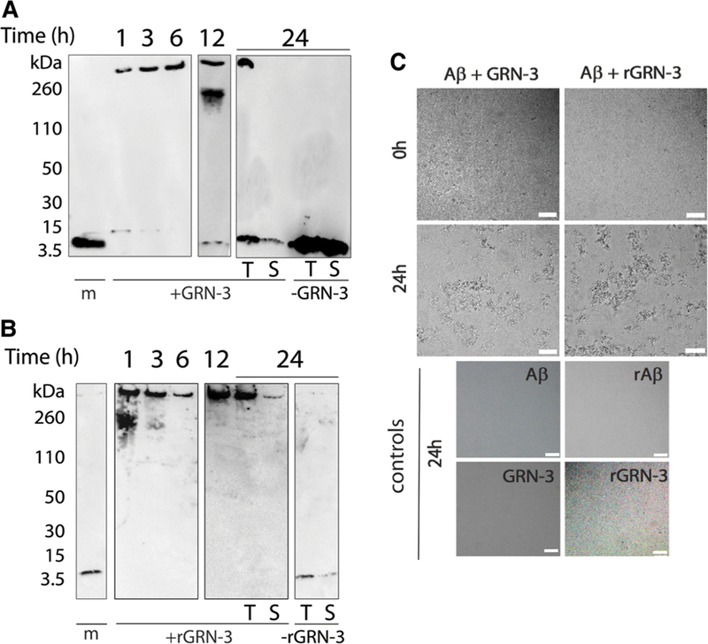
Figure 4. High molecular mass Aβ42 fibrils detected by immunoblots and differential interference contrast light microscopy (DIC).
(A,B) Aliquots of the co-incubated reactions of GRN-3 (A) and rGRN-3 (B), similar to reactions described in Figure 3, were subjected to electrophoresis and immunoblotting at the indicated time points of 1, 3, 6, 12, and 24 h, respectively. Lane ‘m’ represents Aβ42 monomer control. After 24 h, the samples were centrifuged at 18 000×g and both the sample before (total; T) and after centrifugation (supernatant; S) were electrophoresed along with respective controls. (C) DIC was used to observe the fibrillar structures formed by the interaction of Aβ42 with GRN-3. Scale bar denotes 50 μm.

