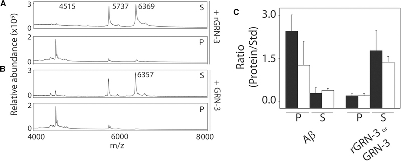
Figure 5. Characterization of fibrils formed upon co-incubation of GRN-3 or rGRN-3 and Aβ42.
MALDI-ToF mass spectra were obtained from the reactions in Figure 4 from both the sedimented pellet and the supernatant after 24 h. Insulin (12.33 μg) was used as an internal standard (Std). (A) MS spectra for the pellet (P) and supernatant (S) from the reaction of Aβ42 (4515 amu) with GRN-3 (6357 amu). (B) MS spectra for the pellet (P) and supernatant (S) from the reaction of Aβ42 (4515 amu) with rGRN-3 (6369 amu). (C) The relative intensities of the proteins in the respective P and S fractions are expressed as a ratio with respect to that of the insulin standard. The closed and open bars represent the reactions with rGRN-3 and GRN-3, respectively.

