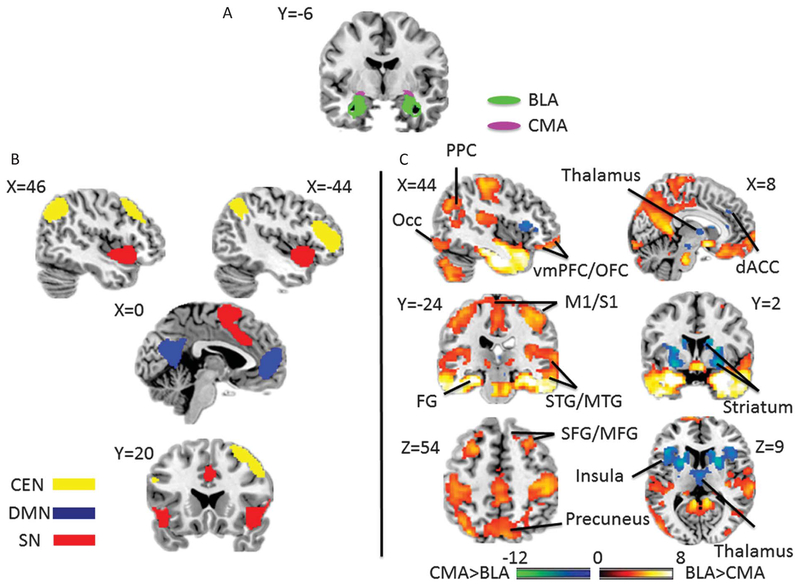
Figure 1.
(A) Seed regions of interest of amygdala subregions; BLA, basolateral amygdala; CMA, centromedial amygdala. (B) Regions of interest of large-scale networks; CEN, frontoparietal “central executive network”; DMN, medial prefrontal-medial parietal “default mode network”; SN, dorsal anterior cingulate–anterior insula “salience network.” (C) Regions of interest of expected targets for amygdala subregions resulting from a voxel-wise 1-way analysis of variance contrasting the BLA and the CMA for the independent NKI sample (false discovery rate (FDR) q < 0.05). Hot color indicates regions having greater connectivity with BLA than CMA, and cool color shows regions connected more with CMA than BLA. dACC, dorsal anterior cingulate cortex; FG, fusiform gyrus; M1/S1, primary somatosensory and motor cortices; Occ indicates occipital cortex; PPC, posterior parietal cortex; STG/MTG, superior temporal gyrus/middle temporal gyrus; SFG/MFG, superior frontal gyrus/middle frontal gyrus; vmPFC/OFC, ventromedial prefrontal cortex/orbitofrontal cortex; and VTA/SN, ventral tegmental area/substantia nigra.

