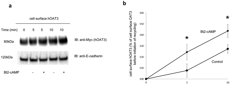
Fig. 4. Biotinylation analysis of Bt2-cAMP-modulated hOAT3 recycling.
(a). Top panel: hOAT3 recycling (5 min and 10 min) was analyzed as described in the section of “Materials and Methods” in the presence and the absence of Bt2-cAMP (10μM), in conjunction with immunoblotting (IB) using anti-Myc antibody (1:100). hOAT3 was tagged with the Myc epitope to facilitate the immunodecetion. Bottom panel: The identical blot as the top panel was re-probed with anti-E-cadherin antibody. E-cadherin is a cell membrane marker protein. (b) Densitometry analyses of results from Fig. 4a, Top panel along with other experiments. Total biotin-labeled hOAT3 was expressed as % of OAT3 biotinylated at 4 °C. Values are mean ± S.D. (n = 3). *P<0.05.

