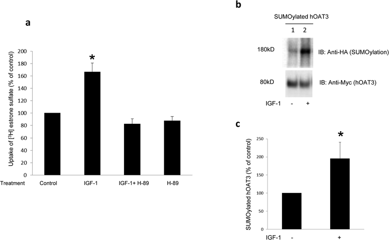
Fig. 9. The effect of IGF-1 on OAT3 transport activity and SUMOylation.
(a) The effect of IGF-1 on hOAT3 transport activity. hOAT3-expressing cells were pretreated with or without a PKA inhibitor H-89 (20μM, 10min). After that, the cells were treated with IGF-1 (100nM, 3hrs) in the presence and absence of PKA inhibitor H-89 (20μM, 3hrs), or H-89 alone, followed by [3H] estrone sulfate uptake (4min, 0.3 μM). Uptake activity was expressed as % of the uptake in control cells. The data correspond to the uptake into hOAT3-expressing cells minus uptake into mock cells and was normalized to protein concentration. Values are mean ± S.D. (n = 3). *P<0.05. (b) The effect of IGF-1 on OAT3 SUMOylation. hOAT3-expressing cells were transfected with HA-SUMO2 and 2.4μg of Ubc9 for 48hrs, then treated with the IGF-1 (100nM, 3hrs). hOAT3 was pulled down by anti-Myc antibody, in conjunction with immunoblotting (IB) using anti-HA antibody. (c) Densitometry analyses of results from Fig. 9b. Values are mean ± S.D. (n = 3). *P<0.05.

