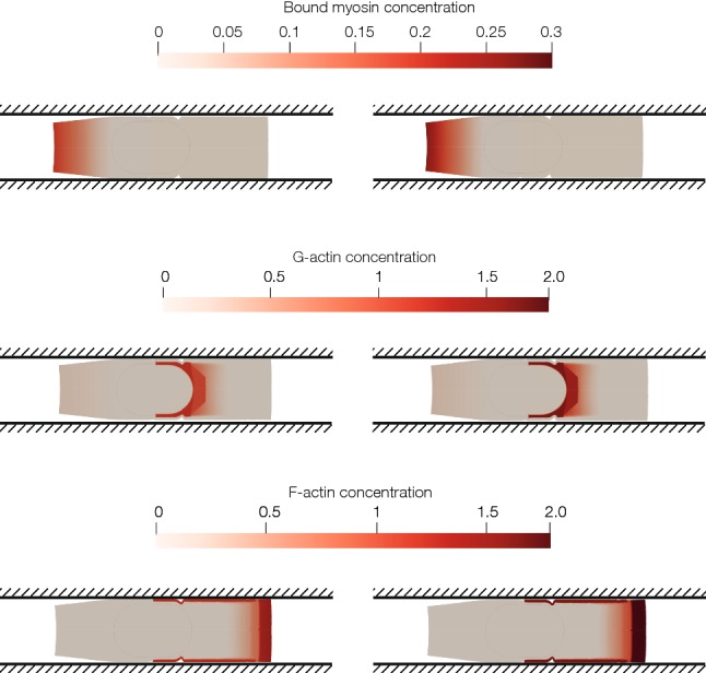
Fig. 5.

Spatiotemporal proteins’ concentration in time 30 min and 50 min. a Concentration of myosin. Myosin is located at the rear part of the cell. Myosin’s concentration depends on the longitudinal coordinate; thus, the lowest value is located near the nucleus and in the front part of the cell. Since the main contraction is generated by myosin attached to the CSK in the rear part of the cell, only this one is modelled. b Concentration of G-actin. Depolymerization takes place near the nucleus. Thus, the highest value of the G-actin concentration is located close to the nucleus. c Concentration of F-actin. The adherent network of F-actin is located in the cell–wall interface. In the cell front, there is a peak of concentration close to the membrane. Concentration depends on the longitudinal distance to the cell nucleus
