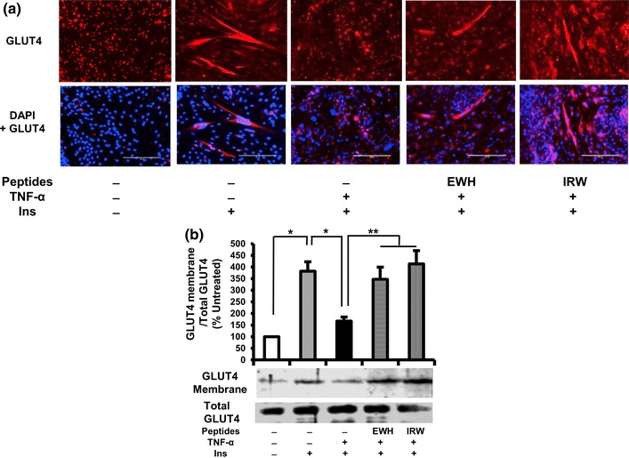
Fig. 3.

Effects of EWH or IRW on GLUT4 translocation in TNF-α-treated L6 myotubes. The myotubes were treated with 5 mg/ml of EWH or 100 µM of IRW for 2 h followed by treatment with 5 ng/ml of TNF-α for 24 h. L6 myotubes were preincubated in KHH buffer for 2 h. They were then incubated in KHH buffer containing 11 mM glucose without or with 100 nm of Insulin for 30 min (a). GLUT4 localization was detected using immunofluorescence technique and western blotting. Cellular localization of GLUT4 proteins is shown in red fluorescence (top) and merged image is also shown (below). A representative set of images from three independent experiments is shown. b Membranes were separated from the cells. The expression of GLUT4 was tested by western blot. Each value represents the mean ± SEM of three independent experiments. Single asterisk indicates p < 0.05 as compared to insulin alone. Double asterisk indicates p < 0.05 as compared to insulin and TNF-α
